I know you are afraid to visit your physician thinking that he might ask you to undergo knee replacement surgery.
Every time you have visited your doctor in the past you been told that you can get your knee pain free only with knee replacement procedure but you have never been advised on trying knee physical therapy rehab first.
You must have taken prescribed countless pain medications to help you with your knee pain but, still it doesn't feel any better and you are looking for ways to get freedom from your knee pain without undergoing knee operative procedures.
I know knee pain is quiet unpleasant and frustrating. But, know what? It's 100% treatable with physical therapy without undergoing painful invasive procedures. Now, let me show you how can you get rid of your knee pain .
First, let's learn about your knee injury and pain so that you can minimize the stress and strain on knee supporting structures and maximize the strength and endurance of the muscles around knee which will tremendously take you one step ahead in alleviating your knee pain and improve performance in daily activities.
")
Knee muscles, ligaments and cartilages
Below you will learn:
Your Knee joint is complex structure formed by .3 bones: Femur (thigh bone), Tibia (Shin bone
")
")
Failure of these ligaments and muscles to provide enough stability along with other movement faults result in various pain conditions of the ankle.
")
1. Knee Arthritis (Joint Inflammation): There are different types of arthritis which affects the knee. the most common ones are osteoarthritis and rheumatoid arthritis
a. Osteoarthritis: This type of arthritis affects weight bearing joints of the body, common in knee and hip joints. Here, knee undergo age related degenerative process as a result of "wear and tear" of joint cartilages. It is more common in people above age of 50.
- Symptoms: Pain, muscle weakness, joint instability and limitation of joint motion, in severe cases it results in knee deformity (bowing of knee joint).
b. Rheumatoid Arthritis: It's an autoimmune disease where person's own immune system attacks multiple joints of the body. Early symptoms of rheumatoid arthritis are manifests first in the hands and feet, knee involvement is seen with disease progression
- Symptoms: Warm and swollen knee joints, limited joint mobility and knee joint deformity is seen with disease progression (knocking knee deformity)
2. Knee Ligament Injuries: Ligaments most frequently occur in individuals between 20-40 years of age as a result of sports injuries (Tennis, basketball, football) but can occur in people of all ages.
There are 4 main ligaments in the knee joint: Anterior cruciate ligament, Posterior Cruciate Ligament, Medial collateral ligament, Lateral collateral ligament.
Cruciate Ligament Injuries: Anterior and posterior cruciate ligament cross each other inside knee joint. Anterior (in front) cruciate ligament prevent excessive forward translation of shin bone whereas posterior (in the back) cruciate ligament restricts excessive backward translation of shin bone to maintain stability in the knee joint.
a. Anterior cruciate ligament injuries (ACL): It is the most common type of knee injury, often results from direct blow on the outer side of the knee or sudden change of direction or speed when running with foot in contact with the ground or extreme forceful over extension of knee.
- Symptoms: Sudden popping of the knee is heard along with swelling and knee feels unstable
b. Posterior Cruciate Ligament Injury (PCL): It most commonly occurs as a result of sudden forceful bending of the knee or intense blow in front of the shin bone with knee bent, example: falling onto a bent knee. Besides, feeling the pain in the back of the knee, typical symptoms of this injury are swelling and stiffness in the knee.
Collateral ligament Injuries: There is a collateral ligament on each side of the knee. Medial collateral ligament on the inner side of the knee and Lateral ligament is present on outer side of the knee.
a. Medical collateral ligament Injuries (MCL): It results from the violent blow on the outer side of the knee. Symptoms: Swelling and pain on the inner side of the knee.
b. Lateral Collateral Ligament Injury (LCL): This type occur as a result of the forceful blow on the inner side of the knee which causes pain and swelling on the outer side of the knee.
3. Meniscal (cartilage) Injuries: There are two 'C" shaped strong cartilages between thigh bone (femur) and shin bone (tibia): medial meniscus (on inner side) and lateral meniscus (on outer side).
Meniscus in the knee help by providing a cushion between thigh bone and shin bone and act as a shock absorber. Medial (inner side) meniscus than lateral (outer side) meniscus.
a. Medial Meniscal Injuries or Tear: It may occur when the foot is fixed and thigh bone rotates internally (towards inner side). Example: while getting out of the car.
b. Lateral Meniscal Injuries or Tear: It results from lateral rotation of the thigh (towards outer side) with foot fixed on floor.
Common symptoms of meniscal injuries or Tear:
- Meniscal tear results in acute locking of the knee or
- Chronic meniscal injuries results in intermittent catching/locking
- Knee pain: In joint line during forced over extension (knee straightening) or full knee bending
- Knee joint swelling and loss of front thigh muscle mass (quadriceps muscle atrophy)
- During catching/locking meniscal injury episode or in chronic cases, knee does not straighten fully
- Unexpected locking and inability to take the load on injured knee while walking
- In case of swelling in meniscal injuries or tear, knee doesn't straighten or bend fully
4. Patellofemoral pain syndrome (PFPS): It is most common in athletes and young adults who are involved in vigorous activities such running, jumping and climbing stairs. It results from knee cap mal-alignment, inner front thigh muscle (VMO), hip abductor and extensor muscles weakness (buttock muscle group) which causes mechanically faulty knee movements and increases the stress over knee cap against thigh bone.
Symptoms includes:
- Aching pain underneath knee cap
- Knee cap crepitational sounds and swelling
- Front knee especially going down the stairs and after prolonged sitting
- You may note popping sounds in the knee while getting up after long sitting hours and climbing stairs
5. Patellar Tendonitis: It refers to the inflammation of tendon that connects knee cap to shin bone. It's common in athletes and young adults who are engaged in frequent explosive knee joint activities such as running or jumping.
Symptoms:
- Starts with dull ache which becomes sharp with increase with impact knee activities
- Patellar tendon inflammation results in weakening of tendon, making it prone to tear
- Patellar Tendon tear results in sharp pain in front of the knee with swelling and feeling of knee instability while walking.
6. Chondromallacia Patella: In this condition the cartilage behind knee cap becomes soft and starts to undergo wear and tear. It is seen in some people with patellofemoral pain syndrome (PFPS). It results from overuse of the knee joint or keeping the knee bent for longer hours
Symptoms are:
- Sharp or aching pain in the knee
- Pain is located in front or around or behind knee cap
- Sometimes presents swelling or crepitational (popping sounds)
7. Iliotibial Band Friction Syndrome (ITBFS): refers to inflammation of iliotibial band, a thick fibrous band that runs along the outer thigh. This band helps in reducing the compressive forces on knee joint during weight bearing activities.
This injury is very common among runners, results pain and inflammation on lower outer thigh (almost 2 cm above outer knee joint line).
The factors which increases stress on Iliotibial band:
a. Downhill walking/running and
b. Increase in distance between feet while walking/running
8. Prepatellar bursitis: It is an inflammation of prepatellar bursa (fluid filled sac over the knee cap). It is most commonly seen in people who expose front of the knee to the ground like in prolonged kneeling position.
Symptoms:
- When inflamed knee movements get restricted due to swelling and pain on direct touch on inflamed area
- Front knee pain due to increased pressure by the knee cap during movement.
9. Osgood Schlatter's Disease: It occurs during adolescence as a result of the overuse of knee joint during rapid growth.
It's self limiting condition and symptoms include pain and swelling below knee cap which increase with activities and improves with rest.
There are number of bio-mechanical faults which results in Knee injuries
- Faulty movement patterns: Foot rolling inwards: Over Pronation, foot rolling outwards: Supination, Leg bone abnormal rotation: tibial torsion places abnormal stress on ligaments and knee cartilages result painful injuries.
- Malalignment or tracking (movement) of the knee cap
- Increase or decrease in Quadriceps angle ("Q" angle)
- Tight Iiliotibial band and ligament structures around knee cap
- Improper foot landing while running and jumping.
- Abnormal rotation of thigh or shin bone.
- Inflexibility of muscles around hip, knee and ankle.
- Muscle imbalances around Knee and hip joint (common pattern involves weakness of inner front thigh muscles and buttock muscles).
- Lack of stability and proprioception (position sense) in the knee joint.
- Right versus left leg difference (Leg Length Difference)
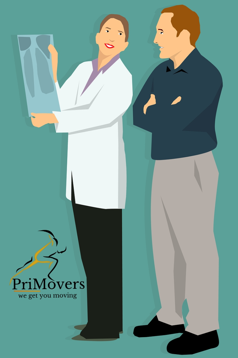
Most knee pain conditions can be diagnosed based on medical history and thorough physical examination.
When discussing about your knee pain try to provide as many details as possible such as location of pain, timing of pain, presenting symptoms, related symptoms, factors which increase or decrease your knee pain. This information will help your movement specialist make the accurate diagnosis.
")
Location of knee pain:
Provide information about the location of your knee pain to your physical therapist or doctor as it provides some very important clues about your problem area, for example:
a. Pain on the outer side of the knee can be due to injury to lateral collateral ligament, iliotibial band, lateral meniscus, whereas
b. Pain on the inner side of the knee indicates injury to medial collateral ligament, medial meniscus, similarly
c. Pain on the back of the knee can result from baker's cyst
Timing of knee pain:
Information about the time of the day when you experience maximum or minimum pain in the knee provides useful insights about your injury. For instance,
1. Knee pain which is more in the morning but improves with day activity indicates inflammatory conditions such as arthritis or iliotibial band friction syndrome (pain in the outer lower thigh)
2. Whether you feel more pain in the knee going upstairs or downstairs. Knee cap related pain (patellofemoral pain) usually felt more on going downstairs and prolonged sitting.
Associated/related symptoms
Do let your physical therapist or doctor know if you experience any other symptoms besides knee pain such sweating, fever or chills, headache, muscle aches, loss of appetite, general body weakness (these are sign of likely infection in the body) or
Full body symptoms like pain and swelling in other joints of the body (hands and feet), fatigue, pins and needles sensation, unexplained weight loss (signs of systemic illness: rhematoid arthritis).
List of physical /clinical tests performed by your physical therapist or doctor.
Knee pain doesn't necessarily have to be from pain source in the knee, it can be from somewhere else such as low back and hip
")
Medical Imaging for Ankle
Based on the impression made by your physical therapist or doctor. They might ask you to get one of the below mentioned investigation test to rule out the issue area in the Ankle. (Only in the required cases)
- X ray: Different views (AP, lateral, oblique) to see any bony changes in case of Osteoarthritis, traumatic arthritis, rheumatoid arthritis,Gout.
- MRI: To visualize soft tissue changes including muscles, ligaments, and joint capsule.
- Diagnostic Ultrasound: It helps in visualizing swelling in the muscle tendons (tendon injuries.
- Nerve Conduction Velocity Test (NCV): In this test needle and surface electrodes are used to find out the intensity of nerve signals transmission to lower body (helps in early detection of nerve compression)
Prognosis of Ankle pain is quite good but it depends on the type and severity/grade (in case of muscle and ligament injury) of Ankle injury. Most ankle injuries take 4-6 weeks of moderate protection to heal completely.
In case of Grade 1 Ankle sprain, it usually takes 2 weeks for the pain, inflammation to subside, Grade 2 (partial ligament tear) takes 4-6 weeks. Even a grade 3 Ankle sprain with complete ligament tear doesn't require surgery but takes little longer time to heal than grade 1 and 2 of ankle (8-12 weeks)
Best Rehab and Recovery Treatment methods for Knee Injuries
Rest: This is first line of treatment in most of the knee pain conditions, especially in case of overuse injuries. It provides time for knee inflammation to subside. Sometimes, it is the thing required to relieve knee pain.
Ice compression: Besides rest, ice pack compression tremendously helps in reducing swelling, inflammation and pain in the knee. For cold compression you can use ice packs or ice bags.
Cold compression should not be directly over the skin and it's application should not exceed more than 10 mins at a stretch to avoid cold burn. You can do cold compression multiple times a day.
Physical Therapy is indispensable component of treatment towards your knee pain.
Physical Therapists are the movement specialists who can help you reduce knee pain and show you exactly how you can prevent common knee injuries by using various strengthening, stability and stretching programs so that you do not have any functional limitations which can hold you back to perform optimally.
Physical Therapy is indispensable component of treatment towards your knee pain.
Your physical therapists use various therapeutic techniques involving soft tissue manipulation, selective stretching and joint mobilization and customized exercise routine to alleviate your knee pain, inflammation, help you regain range of motion, muscle strength, overall balance and knee joint position sense (proprioception) to get you back to pre-injury levels of activity.
They also educate you about gradual increase in essential corrective muscle strength exercises and when to return to sporting activities.
Unless there is knee joint fracture or severely injured knee, you can start physical therapy immediately after injury without any prescription by any doctor.
Apart from rehab specific treatment techniques of mobilizing and stretching, strengthening your muscles around knee joint, your physical therapist might use additional supportive techniques like bracing your ankle, splinting and kinesiology or sports tapping to provide extreme stability to knee to enhance healing and recovery.
Based on your knee pain diagnosis, your physical therapist or doctor might prescribe you knee brace or splint to help keeping your pain and inflammation on check. For instance, ACL or PCL hinge brace in case of Anterior cruciate ligament (ACL) or posterior cruciate ligament (PCL) injuries, patellar tendon straps or supportive tapping in case of patellar tendonitis, soft elastic wraps in case pre-patellar bursitis.
Other treatment options include pain medications, injections and operative procedures.
Pain Medications (NSAIDs): Your doctor might prescribe you pain medications (NSAIDs: Non steriodal Anti-inflammatory drugs) to reduce pain, swelling and inflammation during acute episodes of inflammatory conditions such as bursitis, tendonitis and arthritis.
- Get your knee examined by your physical therapist or physician to know the root cause of your knee pain.
- Discuss with your Physical Therapist to understand drug free and noninvasive treatment methods. Learn various activity modifications and exercises that helps offloading your knee stress.
- Learn the correct exercises by your Physical Therapist as most of the knee pain conditions can be improved with offloading, specific stretching and strength techniques and improving joint movement mechanics.
- Continue with your exercise program given by your physical therapist minimum 3-4 times a week even when you are pain free (as a maintenance program).